

Advanced Diagnosis, Laser Lithotripsy, and Endoscopic Treatment in Nagpur
Kidney stones (renal calculi) are hard mineral and salt deposits that form inside the kidneys. In regions like Vidarbha and Central India—where high ambient temperatures can contribute to dehydration—kidney stone disease is a common cause of acute flank pain and urinary tract obstruction.
Early diagnosis and appropriate clinical intervention prevent severe complications such as urinary tract infections (UTIs), obstructive uropathy, and kidney function impairment.
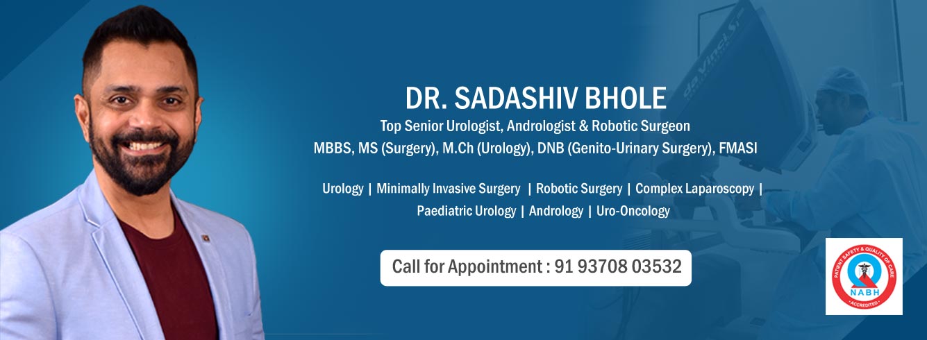
At Ketki Nursing Home & Urology Hospital in Dhantoli, Nagpur, Dr. Sadashiv Bhole provides comprehensive endourological management for kidney, ureteric, and bladder stones utilizing advanced laser and endoscopic techniques.
Common Symptoms of Kidney Stones
Small stones may pass through the urinary tract silently. However, larger or lodged stones cause urinary blockage and trigger distinct clinical symptoms:
- Severe Flank Pain (Renal Colic): Sudden, intense pain in the back and side, below the ribs, often radiating to the lower abdomen and groin.
- Fluctuating Pain Intensity: Pain that comes in waves and fluctuates in intensity as the ureter contracts to move the stone.
- Hematuria: Blood in the urine, giving it a pink, red, or brownish appearance.
- Urinary Changes: Painful urination (dysuria), persistent urge to urinate, or difficulty passing urine.
- Nausea & Vomiting: Caused by shared nerve pathways between the kidneys and the digestive tract.
- Fever & Chills: Secondary sign indicating a potential urinary infection associated with obstruction—a situation requiring urgent urological evaluation.
Diagnostic Evaluation
Precise evaluation determines the exact size, location, density, and anatomical context of the stone, ensuring the selection of the most suitable treatment pathway.
- Non-Contrast CT KUB (Kidney, Ureter, Bladder): The clinical gold standard for stone imaging, offering high resolution to identify stone size, hardness (Hounsfield units), and location.
- Ultrasonography (USG KUB): A quick, radiation-free imaging modality useful for detecting kidney swelling (hydronephrosis) and acoustic stone shadows.
- Laboratory Investigations: Complete blood counts, serum creatinine, blood urea nitrogen (BUN), and urine routine/culture to assess renal function and rule out active infection.
- Metabolic & Stone Analysis: For recurrent stone formers, evaluating urinary pH, serum calcium, uric acid, and retrieved stone composition helps establish preventive strategies.
Modern Treatment Approaches for Kidney Stones
Treatment choices depend on stone size, location, hardness, and patient anatomy. Conservative management is considered for small stones, while advanced endoscopic and laser procedures are used for obstructive or larger stone burdens.
Conservative & Medical Management
Small stones (under 5 mm) without infection or significant kidney swelling may pass spontaneously with increased fluid intake, analgesics for pain relief, and Medical Expulsive Therapy (MET) using medications to relax ureteric smooth muscle.
Endoscopic & Laser Surgical Interventions
When intervention is required, minimally invasive endourology allows for complete stone fragmentation and clearance through natural pathways or minimal keyhole access:
- RIRS (Retrograde Intrarenal Surgery):
- Mechanism: A flexible ureteroscope is advanced through the natural urinary passage into the kidney’s internal collecting system. High-precision laser energy (such as the Thulium Fiber Laser system) is used to dust or fragment the stone into tiny particles.
- Advantage: No external skin incisions, minimal tissue trauma, and rapid recovery.
- PCNL (Percutaneous Nephrolithotomy):
- Mechanism: A minimal keyhole access tract (approx. 1 cm) is established directly through the back into the kidney under C-arm fluoroscopic guidance. Rigid or flexible nephroscopes are inserted to fragment and extract stone pieces.
- Indication: The established treatment choice for large stones (greater than 2 cm), staghorn calculi, or complex stone burdens.
- Advantage: High single-session stone clearance rate for large stone volumes.
- Ureteroscopy (URS):
- Mechanism: A thin, rigid or semi-rigid ureteroscope is passed into the ureter to visualize and break ureteric stones using laser energy or pneumatic lithotripters, followed by basket retrieval.
- Indication: Direct clearance of mid- and lower-ureteric stones causing acute pain or obstruction.
- Advantage: Immediate relief of acute obstruction and severe pain.
Kidney stones are hard mineral deposits that form in the urinary tract. They can cause pain, bleeding, obstruction and, when associated with infection, require urgent medical evaluation. At Ketki Nursing Home & Urology Hospital in Dhantoli, Nagpur, Dr. Sadashiv Bhole provides evaluation and endourological treatment for kidney and ureteric stone disease using appropriate laser and endoscopic techniques.
Clinical Experience in Endourology
Surgical treatment of complex stone disease relies on procedural precision and clinical judgment. Dr. Sadashiv Bhole brings over 31 years of clinical experience and a track record of 65,000+ career surgeries, including more than 42,000 complex and advanced urological procedures.
Institutional care is provided at Ketki Nursing Home & Urology Hospital in Dhantoli, Nagpur, a dedicated 25-bed urological center with 25 years of institutional history and 35,000+ institutional urological procedures. The hospital’s supplied equipment inventory includes Thulium Fiber Laser systems, Olympus and WOLF nephroscopes/ureteroscopes and high-definition intraoperative imaging.
Preventive Strategies for Stone Recurrence
Preventing recurrent stone formation is an integral part of urological care:
- Hydration: Fluid intake recommendations should be individualized according to stone type, kidney function, comorbidities and clinician advice.
- Dietary Adjustments: Balanced sodium intake, moderate animal protein consumption, and appropriate dietary calcium intake based on metabolic evaluation.
- Regular Monitoring: Periodic ultrasound or clinical reviews for individuals with a history of recurrent stone formation.